Nerves
Nerves are everywhere and therefore are affected by almost all of these procedures. Even in aneural tissues (ex tendons and ligaments) after injury they too become innervated and pain sensitive.
Nerves are very dependent on an abundant blood supply and when under stress produce proteoglycans.
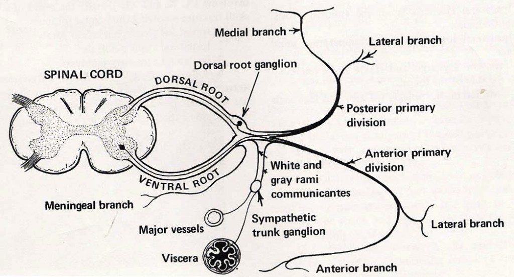
This section will deal with stretching the long nerves of the primary ventral rami (these form the plexuses) and those of the primary dorsal rami. The cranial nerves will in the head and neck section. For the most part, the autonomics will be discussed as parts of other sections.
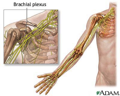
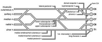
- Brachial plexus – RN, UN, MN, Musculocutaneous, branches to rotator cuff, latissimus, pectoralis, serratus anterior.
- Cervical plexus – This will be covered in the head and neck video.
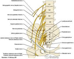
- Lumbar plexus – Obturator, Femoral
- Lumbosacral plexus – Common peroneal, Tibial, Nerves to the buttocks
- Cervical spine – Dorsal primary rami will be covered in the head and neck
- Lumbar spine – Dorsal primary rami
General comments:
- These nerves generally follow muscles and therefore the various techniques are similar to those of the muscles, tendons and entheses.
- These nerves generally follow blood vessels and so will be similar to blood vessel stretching techniques.
- Distal aspects of the nerves are involved in virtually any procedure you do. For example, nerves in the cortex of the bone will be loaded when that bone is flexed. Nervi nervorum are an example of the distal end of small nerves supplying large nerves especially proximally.
- Obviously increased pressure and a lack of blood supply will led to neuropathic pain. This is especially true for those nerves derived from post traumatic neurogenesis.
Videos
Large Nerves
All nerves have a blood supply. This includes nervi nervorum. Degeneration and old injury may have led to neurogenesis. It’s my opinion non vascular edema may exist at these sites and cause neuropathic pain. End range loading these large nerves and plexi will result in an increased blood supply.


Videos
Dorsal Primary Rami
All the muscles, ligaments and fascia of the back are innervated by nerves of the dorsal primary rami. The zygapophyseal joints along with their capsuls and ligaments are also included as well as most of the skin and posterior aspects of the vetebrae. For the most part these nerves are much shorter than their ventral counterparts.

